Reference
Glossary
33 structures and mechanisms of the body and the brain that appear across the site, two lines each, written from the point of view of someone designing, not of a textbook.
Throughout the site, the first mention of any of these terms opens its definition right there, without taking you out of the paragraph. This page is for the other direction: reading them all at once, or going back to one you have seen.
A
- A1
-
The first area to receive sound, in the temporal lobe. It receives everything that comes in through the ears, but only one stream at a time gets priority and reaches consciousness.
Go deeper
It sits in the temporal lobe, above the ear, inside a fold. The path to it is long: sound becomes vibration in the eardrum, becomes movement of fluid in the cochlea, becomes an electrical impulse, climbs through the brainstem and only then reaches the cortex.
A1 is organised by frequency, like a keyboard: low notes on one side, high notes on the other, in order. That organisation is anatomical and was there before you learned anything about sound.
The detail that interests whoever designs is speed: the auditory path has fewer stops than the visual one, and so sound reaches the brain before the corresponding image. It is the reason a sound alert is more effective at interrupting than a visual one, and more aggressive for the same reason.
- amygdala
-
Two small structures in the temporal lobe that decide, in milliseconds, whether something is a threat or an opportunity. They respond to your screen before any conscious judgement about it.
Go deeper
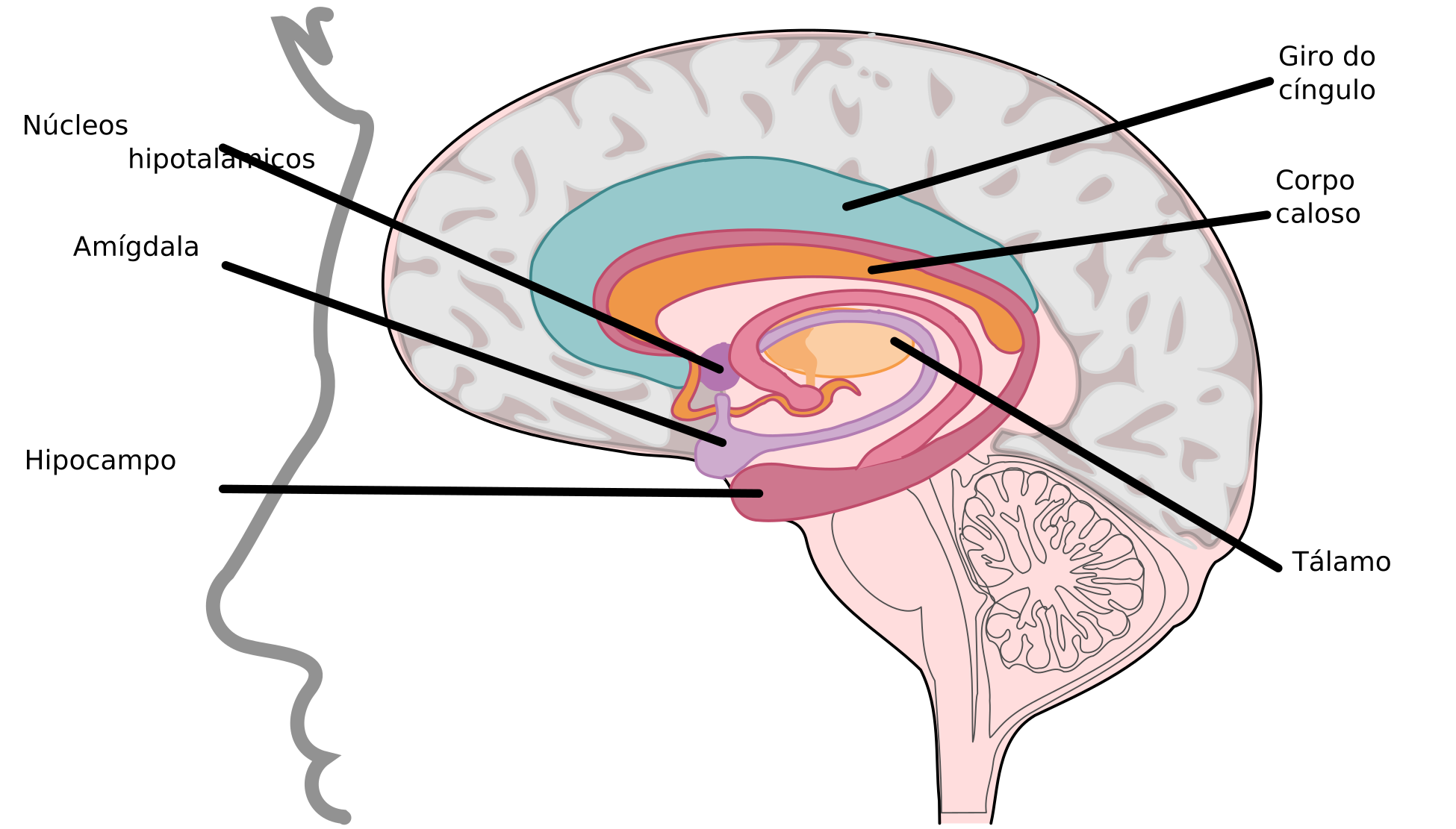
The limbic lobe, in sagittal section
OpenStax College; vectorised by Marnanel · Anatomy & Physiology · CC BY 3.0 · labels translated into PortugueseTwo almond-sized structures, one on each side, at the inner tip of the temporal lobe. That is where the name comes from.
It is the brain’s relevance detector, and it is fast: it receives a coarse version of the sensory signal through a shortcut that bypasses the cortex, and fires before the person knows what they saw. Fear is the most studied response, but it responds to anything that matters, including good things.
In an interface, it is what reacts to the error in red, to the deletion warning, to the countdown. The reaction arrives before the reading, and no later explanation undoes it, only stops it from happening again. That is why the tone of an error message matters more than its content.
Appears in: Negativity bias
B
- basal ganglia
-
Together with the cerebellum, they store what the body learned to do without thinking: typing, swiping, reaching an icon. It is the memory that survives distraction.
Go deeper
A set of nuclei deep in the brain, below the cortex.
It is where repeated behaviour becomes automatic. While a sequence is being learned, it consumes prefrontal cortex; once repeated enough, it migrates here and starts to run at almost no cost, and almost without awareness.
It is the structure behind every habit of use: the path the person takes through your product without looking, the button they hit from muscle memory. It is also why moving an element’s position hurts more than it seems: the automatism does not update on notice, only through new repetition.
Appears in: Clustering illusion, Flexibility and efficiency of use
- brainstem
-
The oldest part of the brain, at the junction with the spinal cord. It handles what is non-negotiable — breathing, waking, reacting to a fright — and it is where almost all the signals from the senses pass through.
Go deeper
The stalk that links the brain to the spinal cord, at the base of the skull. It is the oldest part and the one that cannot be lost: it controls breathing, heartbeat and level of consciousness.
Every auditory signal passes through here before climbing, and it is here that the difference in the arrival of sound between one ear and the other — a matter of microseconds — becomes the perception that something came from the left or from the right.
It is also the origin of the systems that regulate alertness and sleep, which makes it the point where tiredness and attention meet physically. No interface talks to it directly, but every interface is used by someone whose brainstem has already decided how much wakefulness is left for the day.
C
- cerebellum
-
The structure low at the back of the skull that automates movement and sequence. The more someone uses your product, the more the operation migrates here, and the costlier it gets to change what has already become automatic.
Go deeper
The wrinkled structure at the back and bottom of the skull. It takes up about ten per cent of the brain’s volume and contains more than half of all its neurons.
It handles the timing and precision of movement, and, according to the more recent literature, also the timing and precision of operations that are not motor. It anticipates: it predicts where the hand will land and corrects before the error happens.
It is what makes a keyboard shortcut faster than a menu, and what makes a well-calibrated drag feel natural. It is also what suffers when the interface’s response is irregular: a delay that varies prevents prediction, and the gesture stops becoming automatic.
Appears in: Fitts’s law
- cortisol
-
The stress hormone, and a slow hormone: it takes twenty to forty minutes to peak, so it is not what explains the irritation with a frozen screen, but rather what builds up over a whole day of bad work. Sustained, it brings down working memory.
Go deeper
A hormone, not a neurotransmitter: it is released by the adrenal glands and circulates in the blood, which makes it slower and longer-lasting than a neural signal.
It is the hormone of the prolonged stress response. In a short peak, it improves attention and memory. Sustained, it does the opposite: it harms the hippocampus, degrades the formation of new memory and narrows attention to what looks like a threat.
An interface that produces constant pressure — a flashing deadline, a balance in red, a warning that will not close — is not keeping the person alert. It is making them worse at what they came to do, and charging the difference to their memory.
D
- dopamine
-
The chemical signal of reward and anticipation. It is not the molecule of pleasure, but of “worth seeking”, which is why expectation usually pulls more behaviour than delivery does.
Go deeper
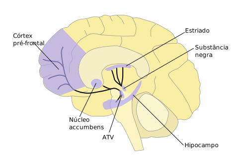
The dopaminergic pathways and the reward circuit
NIDA — National Institute on Drug Abuse; derivative work by Quasihuman · Dopamine pathways · Public domain · labels translated into PortugueseA neurotransmitter, that is, a molecule one neuron releases to talk to another. It is not a structure, it is a chemical signal.
The popular association with pleasure is wrong and worth undoing. Dopamine encodes reward prediction error: it rises when something is better than expected, falls when it is worse, and stays neutral when it is exactly as predicted. It moves you towards, more than it makes you like.
That is why the anticipation of a notification is stronger than its content, and why “dopamine” has become a loose word in product conversation. When someone says a feature “releases dopamine”, the useful question is: compared to what expectation?
- dorsolateral prefrontal cortex
-
The part of the prefrontal cortex linked to self-control and to decision under uncertainty. It is the one that loses to impulse when the immediate reward is visible on the screen.
Go deeper
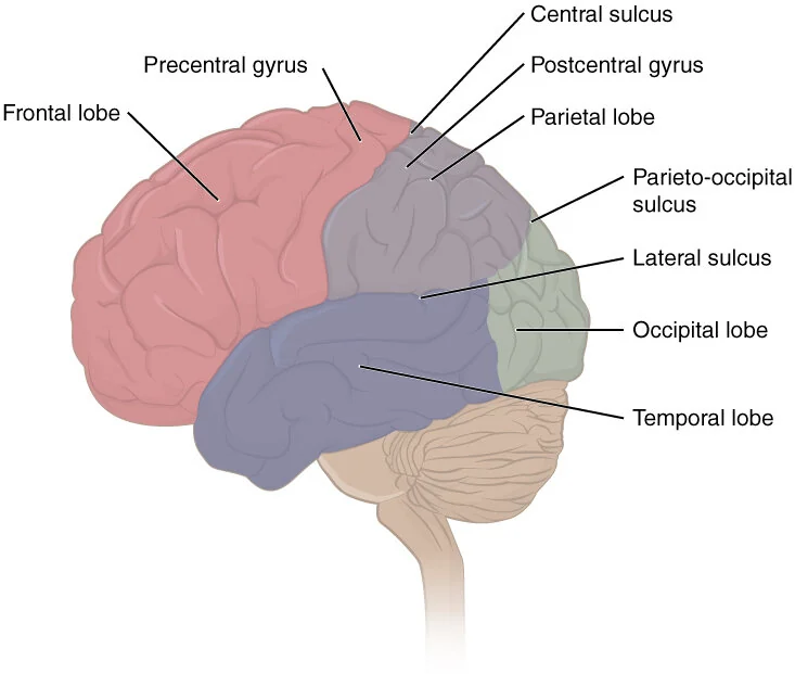
The lobes of the cerebral cortex
frontal (planning and decision) · parietal (space and attention) · temporal (hearing, language and memory) · occipital (vision)
OpenStax · Anatomy and Physiology (2016) · CC BY 4.0The upper, outer part of the prefrontal cortex, on the side of the forehead.
It is the region most associated with holding information active while doing something else: keeping the goal in view, resisting distraction, comparing what is on the screen with what was on the previous screen. It works together with the frontoparietal circuits, and it is one of the first to fail when the task tightens.
It is the structure behind the practical limit this site keeps talking about: whatever the interface asks the person to remember between one screen and the next is charged here, and the balance is low.
E
- echoic memory
-
The sound equivalent of iconic memory: an audio pattern stays available for about three seconds. It is the reason it is possible to answer “what?” and understand the sentence before the person repeats it.
Go deeper
The auditory equivalent of iconic memory, and more generous: the echo of what was said lasts a few seconds, not fractions.
It is the reason you can answer “what?” and, halfway through the question, have already understood, the sentence was still available for internal re-reading.
The difference in duration between the two channels has a practical consequence: spoken instruction has a wider window for recovery than instruction shown and withdrawn. An alert that appears and vanishes depends on the person looking; a sound does not.
F
- frontoparietal circuits
-
The pair that supports working memory: the prefrontal cortex holds the information active while the parietal lobe keeps track of where things are.
Go deeper
The lobes of the cerebral cortex
frontal (planning and decision) · parietal (space and attention) · temporal (hearing, language and memory) · occipital (vision)
OpenStax · Anatomy and Physiology (2016) · CC BY 4.0It is not a structure, it is a conversation: the frontal and parietal regions working in sync, linked by long bundles of fibres.
It is this network that supports working memory and directed attention. It keeps active what matters and suppresses what does not, and it does so at a high metabolic cost, which is why the feeling of mental effort is literal, and not a figure of speech.
Every time a screen forces the person to keep something in their head — a code, a comparison, an instruction that vanished — it is this network that is paying, and it does not scale.
Appears in: Hick–Hyman law, Cognitive load theory, Miller’s law, Salience bias, Flow state
- fusiform gyrus
-
The area that recognises faces and shapes already seen. The more times it meets the same thing, the less effort it spends, and the brain reads that lesser effort as “I like this”.
Go deeper
The lobes of the cerebral cortex
frontal (planning and decision) · parietal (space and attention) · temporal (hearing, language and memory) · occipital (vision)
OpenStax · Anatomy and Physiology (2016) · CC BY 4.0A fold on the underside of the temporal lobe, on both sides.
It contains the most face-specialised region in the whole cortex, and “specialised” here is a strong word: lesions in this area produce prosopagnosia, the inability to recognise familiar faces with vision preserved in everything else. The same region takes part in recognising written words, which suggests that reading is, in part, recognising the shape of the word the way one recognises a face.
That is why faces in an interface capture the gaze before any other element, and why familiar words are read as a block while unknown ones are spelled out. An unfamiliar typeface and all caps undo that shape and charge the difference.
H
- hippocampus
-
The structure that turns recent experience into lasting memory. Everything your interface asks the person to remember tomorrow passes through it today.
Go deeper
The limbic lobe, in sagittal section
OpenStax College; vectorised by Marnanel · Anatomy & Physiology · CC BY 3.0 · labels translated into PortugueseA curved structure on each side of the brain, on the inner part of the temporal lobe. The name comes from the shape, which resembles a seahorse.
It is not where memories are kept: it is what decides what deserves to be kept and makes the transfer. New memory passes through here; old memory already lives spread across the cortex. The best-known clinical case in neuroscience, the patient H.M., lost the hippocampus on both sides in surgery and went on to live in a continuous present: he kept everything he had learned before and formed no new declarative memory.
For whoever designs, the consequence is harsh: nothing your interface teaches is retained for free. What is not repeated, contextualised or unnecessary to remember simply does not survive the session, and a good part of what it teaches today is already gone tomorrow.
Appears in: Jakob’s law, Availability heuristic, Peak–end rule, Von Restorff effect, Serial position effect +1
I
- iconic memory
-
The trace an image leaves for about 250 milliseconds after it leaves the screen. It is what allows comparing two things in the blink of an eye, and what is lost when an element disappears too fast.
Go deeper
The visual trace that survives for a fraction of a second after the image is gone. It is why you can mentally “re-read” what just flashed on the screen.
It lasts around a few hundred milliseconds, and holds much more than the person can report. The classic experiments show exactly that: someone who sees a matrix of letters for an instant can name any row asked for right afterwards, but not all of them, the information was there and faded during the report.
It is the reason very fast animations are perceived without being understood: the signal existed, but did not last long enough to become something else.
L
- limbic system
-
An inherited name for a group of structures — amygdala, hippocampus, neighbours — that the design literature still treats as a single block. Neuroscience does not treat it that way, because there is no anatomical boundary and no common function, and each of these structures does different things.
Go deeper
The limbic lobe, in sagittal section
OpenStax College; vectorised by Marnanel · Anatomy & Physiology · CC BY 3.0 · labels translated into PortugueseA set of structures in the core of the brain — amygdala, hippocampus, hypothalamus and neighbours — historically grouped under the name “emotional brain”.
The term is older than the evidence that supports it, and today it is used with a caveat: the structures exist and talk to one another, but they do not form a system separate from the rest, and emotion does not live in one region. Worth knowing before treating “limbic” as a synonym for irrational.
What survives of the idea is useful all the same: there is a fast circuit, tied to memory and to value, that responds before the slow, deliberate circuit. Designing while ignoring the first is designing for a person who does not exist.
- long-term memory
-
What the person still knows tomorrow of what they saw today. It does not live in one place only: it forms through the hippocampus and, over time, comes to live spread across the cortex.
Go deeper
It is the memory that survives the session. It includes what the person can declare, facts and episodes, and what they know how to do without declaring, like the path to a menu they have used a hundred times.
Its formation passes through the hippocampus and the medial temporal lobe, and the long-term archive is the neocortex. The dependence on the hippocampus decreases over time, as Squire and Zola-Morgan described in 1991.
For whoever designs, it is the side of memory that the interface does not control in the moment. What it teaches today only stays if it is repeated, contextualised or recognisable tomorrow.
Appears in: Jakob’s law, Availability heuristic, Serial position effect
M
- medial prefrontal cortex
-
The part of the prefrontal cortex that thinks about people: what others want, what they think of us, whether they can be trusted.
Go deeper
The lobes of the cerebral cortex
frontal (planning and decision) · parietal (space and attention) · temporal (hearing, language and memory) · occipital (vision)
OpenStax · Anatomy and Physiology (2016) · CC BY 4.0The part of the prefrontal cortex that sits on the inner wall, between the two hemispheres.
It activates when the person thinks about themselves and when they think about other people, and that overlap is no accident of measurement: thinking about what the other person wants uses, in large part, the same machine as thinking about what I want. It is one of the pieces of the network that supports reading other people’s intentions.
It is the substrate of everything in design that depends on “what will they think of me”: social proof, counters of people looking, ratings. It works because the question about the other and the question about oneself run in the same place.
Appears in: Illusion of transparency
- medial temporal cortex
-
The inner part of the temporal lobe, hippocampus and surroundings, through which fact and episode pass to become lasting memory. Its role is temporary: over time the memory ends up stored in the cortex and stops depending on this region.
Go deeper
The inner part of the temporal lobe, which includes the hippocampus and the cortex around it.
It is the set that supports declarative memory, what can be put into words. It works in functional opposition to the basal ganglia, which support what one knows how to do without being able to explain.
The distinction matters in an interface: teaching by text and teaching by repetition record in different systems. The tutorial the person read lives in one; the gesture they repeated a hundred times lives in another, and it is the second that survives a change of layout, which explains why redesigns break precisely the oldest users.
Appears in: Law of visibility, Availability heuristic, Peak–end rule, Von Restorff effect
- mentalising network
-
The circuit that infers what goes on in other people’s heads: intentions, beliefs, desires. It is what comes into play when the interface has people on the other side, in the rating, in the comment, in the count of those who have already bought.
Go deeper
A set of regions — medial prefrontal, temporoparietal junction and neighbours — that activate when the person thinks about what another person is thinking.
It is the theory of mind system: attributing intention, predicting reaction, imagining what the other knows and does not know. It works on its own, without conscious effort, and it is one of the brain’s default modes at rest.
In design, it is what makes the person attribute intention to your product. A screen that hides the cancel button is not read as badly designed: it is read as wanting something. The network does not distinguish interface from person, and the suspicion, once formed, contaminates the rest.
Appears in: Fundamental attribution error
- mirror neurons
-
Cells that fire both when you do something and when you see someone else doing it. They were proposed as the basis of imitation and empathy, a reading the field itself has already corrected, and what is left is that seeing someone act partly activates the system of the one who acts.
Go deeper
Neurons that fire both when the individual performs an action and when they observe another performing the same action. Discovered in monkeys, in the 1990s, by accident.
The existence of the phenomenon is solid; the extent of what it explains is not. The popular reading — that they would be the basis of empathy and culture — went far beyond what the data support, and the field went through a strong correction. Worth citing with care.
What remains applicable is modest and real: seeing someone do partly activates the system of the one who does. It is one of the reasons a video demonstration teaches better than a written description, and an interface that shows the gesture is learned faster than one that describes it.
N
- nucleus accumbens
-
The centre of the reward circuit. When something goes right — a button that responds, a task that closes — it is what records “worth it, do it again”.
Go deeper
The dopaminergic pathways and the reward circuit
NIDA — National Institute on Drug Abuse; derivative work by Quasihuman · Dopamine pathways · Public domain · labels translated into PortugueseA small structure at the base of the brain, at the meeting point between the emotional circuit and the motor one.
It is the centre of the reward system, and the finding that changed everything is that it responds more to expectation than to the gain. The response is at its peak when the reward is possible and uncertain; when it is guaranteed, the response drops.
It is the neural basis of the infinite feed, the loot box and “pull to refresh”. Uncertainty is not a side effect of these patterns: it is the mechanism. Knowing that is what separates using it to give a task rhythm from using it to hold someone captive.
O
- orbitofrontal cortex
-
It sits right behind the eyes and assigns value: whether the thing is worth it, whether it is pleasant, how much it costs in effort. It answers before the person knows they asked.
Go deeper
The lobes of the cerebral cortex
frontal (planning and decision) · parietal (space and attention) · temporal (hearing, language and memory) · occipital (vision)
OpenStax · Anatomy and Physiology (2016) · CC BY 4.0It sits just above the eye sockets, at the base of the frontal lobe.
It is where value is assigned. Not the price, the subjective value, the one that changes with context, expectation and what came before. The same offer is worth more after a high anchor and less after a low one, and this is where that recalibration happens.
Anchoring, loss aversion and the endowment effect depend on this region. It is also why these patterns are so effective and so easy to turn into manipulation: they do not convince anyone of anything, they just change the ruler the person measures with.
Appears in: Endowment effect
- otoliths
-
Two pouches with calcium crystals on a membrane, also in the inner ear. They register acceleration in a straight line and the direction of gravity, which way is down.
Go deeper
While the semicircular canals handle rotation, the otoliths handle straight-line displacement and gravity. They are two pouches, the utricle and the saccule, with a gelatinous membrane covered in calcium carbonate crystals.
The crystals are denser than the rest and, because of that, lag behind when the body accelerates and slide when the head tilts. Their displacement bends the cilia underneath. That is how you know the lift has started going up with your eyes closed, and that is how you know which way the floor is.
They do not distinguish acceleration from tilt, because both produce the same force on the membrane. What resolves the ambiguity is vision, crossing the two pieces of information, and it is in that stitching that motion sickness lives.
P
- place and grid cells
-
The internal GPS, in the hippocampus and its surroundings. It evolved to remember paths to water and food, and it is the same one that remembers where that button was on the previous screen.
Go deeper
Neurons in the hippocampus and neighbouring regions that fire when the animal is at a specific point in space. Grid cells do the same in a hexagonal pattern, like internal graph paper. The discovery earned the 2014 Nobel Prize.
What came afterwards is what matters here: the same machinery seems to organise not only physical space but abstract relations, the proximity between ideas, the position of an item in a sequence, the path travelled through a subject.
It is the most likely neural basis of the metaphor design uses all the time without thinking: “where am I”, “back”, “go deeper”. The person is not imagining a place out of poetic licence; they are using the system that evolved for places.
- prefrontal cortex
-
The region behind the forehead, where the person decides, plans and sustains attention. It keeps one thing in focus at a time and inhibits the rest: it is where the bottleneck of conscious decision usually tightens.
Go deeper
The lobes of the cerebral cortex
frontal (planning and decision) · parietal (space and attention) · temporal (hearing, language and memory) · occipital (vision)
OpenStax · Anatomy and Physiology (2016) · CC BY 4.0It is the region behind the forehead, and it is the last part of the brain to finish maturing, a process that runs until around the age of twenty-five. It is also the first to lose efficiency under tiredness, hunger, stress or alcohol.
It is where the person holds a goal while doing something else, compares alternatives, inhibits the immediate impulse and picks up where they left off after an interruption. None of this is automatic: all of it costs, and the budget is small and runs out.
Most of what this site calls “load” happens here. Every extra field in the form, every decision the interface hands over to whoever uses it, every instruction that has to stay in the head instead of on the screen: all of it is charged to this region, which arrives at your product already partly spent by the rest of the day.
Appears in: Cognitive load theory, Miller’s law, Clustering illusion, Choice-supportive bias, Flexibility and efficiency of use
S
- S1
-
The map of the body drawn over the cortex. Fingers, lips and tongue take up enormous areas; back and legs, almost nothing: that is why the fingertip tells apart what the elbow cannot.
Go deeper
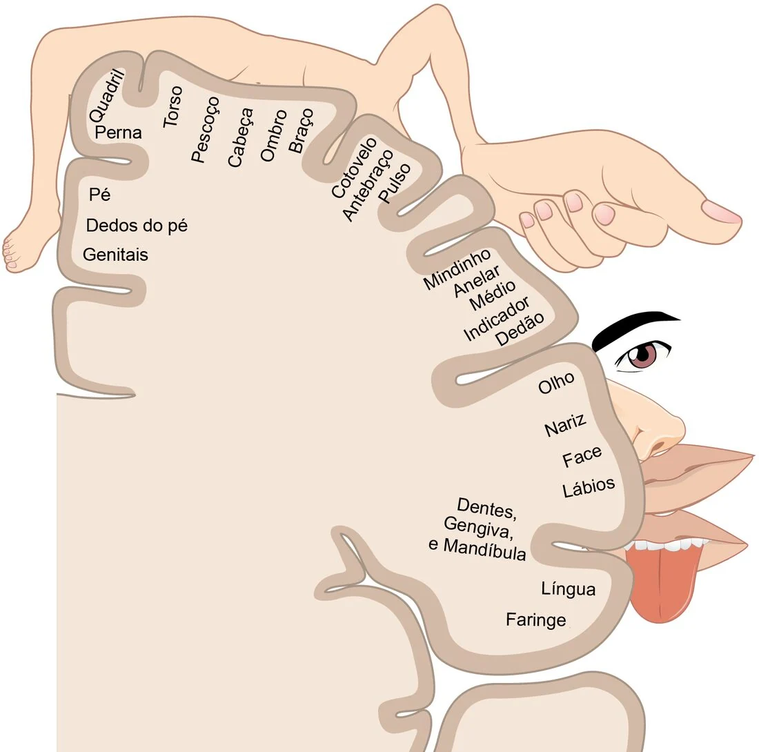
The sensory homunculus of the primary somatosensory cortex (S1)
OpenStax College; derivative work by Ederporto · Anatomy & Physiology (Portuguese edition) · CC BY 3.0It is a strip that crosses the top of the head from ear to ear, just behind the strip that commands movement. Each piece of it corresponds to a piece of the body, in order.
The order is preserved, but the proportion is not: the map is drawn by number of sensors, not by the size of the part. Lips, tongue and fingertips take up enormous areas; the back and the thighs take up almost nothing. It is this distorted map that the figure alongside draws.
That is why touch interaction works: the fingertip is one of the regions of highest tactile resolution in the body. And that is why tactile feedback — the short vibration when confirming an action — is perceived even when the eye is elsewhere.
- semicircular canals
-
Three rings of fluid in the inner ear, one for each axis of rotation. When the head turns, the fluid takes a moment to follow, and it is that delay the body reads as “I turned”.
Go deeper
They are three ring-shaped tubes, filled with fluid, mounted at nearly perpendicular angles to one another: one for each axis of head rotation. Inside each one there is a gelatinous gate with hair cells.
When you turn your head, the bone turns with it and the fluid lags behind for an instant, by inertia. That drag pushes the gate, bends the cilia and changes the firing rate of the fibre. That is why they detect rotational acceleration, and not constant rotation: in a chair spinning at a fixed speed, the fluid catches up with the bone and the sensation of spinning disappears.
For whoever designs, what matters is what they do not do: they do not know you are looking at a screen. They report what the skull did, and nothing more.
- superior colliculus
-
A station in the brainstem that pulls the gaze towards what moves or contrasts, before the person decides to look. It is that reflex that every visual highlight hijacks.
Go deeper
A small structure in the brainstem, very old in evolutionary terms, earlier than the visual cortex.
It receives a copy of the visual signal through a shortcut that bypasses the cortex, and commands the reflex movement of the eyes towards what moved or appeared suddenly. It is the reason you are already looking at the thing before you know what it is.
It is the structure a flashing banner hijacks. And it is the reason that kind of device works even on those who hate it: the shift of gaze does not pass through your opinion of it.
Appears in: Salience bias
T
- thalamus
-
The brain’s distribution hub: almost everything that comes from the senses passes through it before reaching the cortex. Smell is the odd one out, with a direct route to the areas of emotion and memory, and that is where the feeling-laden memory a scent pulls up comes from.
Go deeper
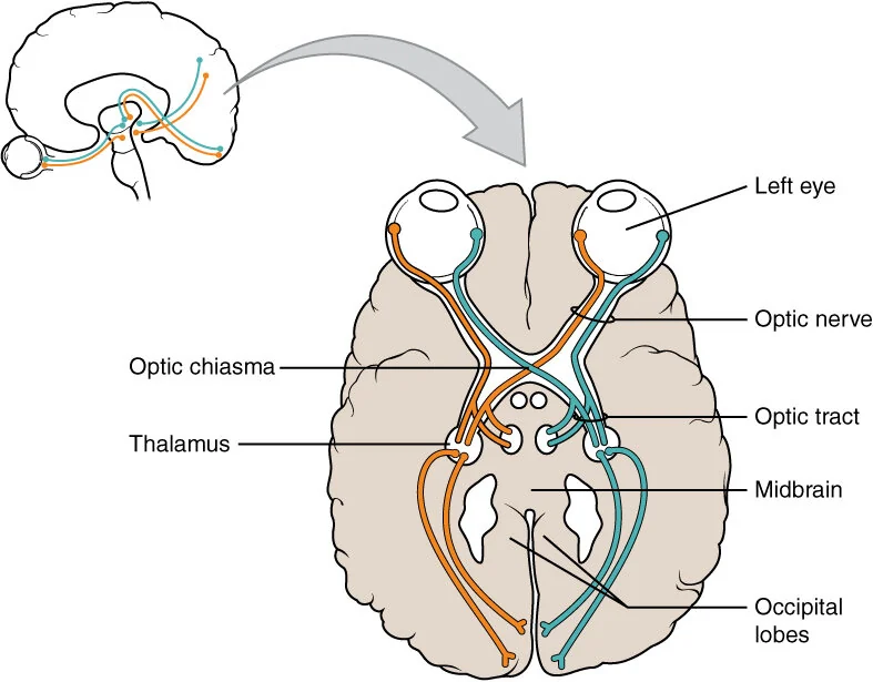
The visual pathway, seen from below
eye → optic nerve → optic chiasm → optic tract → thalamus (lateral geniculate nucleus) → occipital lobe, where area V1 sits
OpenStax · Anatomy and Physiology (2016) · CC BY 4.0It sits at the geometric centre of the brain, roughly where you would point if you had to point to the middle of your own head. It is a paired structure: there is one on each side.
Almost everything the senses capture passes through here before reaching the cortex, the known exception being smell, which comes in through another door. The thalamus is not a pass-through cable: it regulates what passes and with what intensity, and it is one of the pieces that decide what gains access to consciousness when there is more going on than fits.
It is the structure that explains why attention is subtraction, not addition. There is no such thing as “paying more attention to everything”: what comes in extra on one channel comes in less on another.
V
- V1
-
The first area of the brain, right at the back of the head, to receive what the eye sent. It is where the image becomes contrast, edge and orientation, before you recognise what you are looking at.
Go deeper
The visual pathway, seen from below
eye → optic nerve → optic chiasm → optic tract → thalamus (lateral geniculate nucleus) → occipital lobe, where area V1 sits
OpenStax · Anatomy and Physiology (2016) · CC BY 4.0It sits at the back of the head, at the rear pole of the brain, as far from the eyes as possible, which already says something: the signal crosses the entire head before it becomes an image. It leaves the eye through the optic nerve, passes through the thalamus and arrives here.
What V1 does is not seeing: it is decomposing. Each group of neurons responds to something very specific: an edge tilted at 30 degrees, a light–dark contrast, a movement to the right. None of them knows what the object is. The image you think you are seeing does not yet exist at this stage.
For whoever designs, what matters is what V1 delivers for free and what it does not: contrast, edge and orientation are processed before any decision of yours about where to look. That is why an element with a strong edge is found without effort, and why low-contrast text costs dearly even when it is legible.
Appears in: Law of proximity, Law of similarity, Law of uniform connectedness, Law of Prägnanz, Law of legibility
- V2
-
The area right after V1. It joins the edges V1 detected, separates figure from ground and decides which elements form a group, all of it before you notice you looked.
Go deeper
The visual pathway, seen from below
eye → optic nerve → optic chiasm → optic tract → thalamus (lateral geniculate nucleus) → occipital lobe, where area V1 sits
OpenStax · Anatomy and Physiology (2016) · CC BY 4.0It sits right around V1, like a ring. It receives the loose edges from it and does the first assembly.
This is where an edge becomes the outline of something. V2 decides what is figure and what is ground, closes shapes that are partly hidden and groups elements that belong together. The Gestalt laws — proximity, similarity, closure — describe the behaviour of this stage, and that is why they seem obvious: you do not choose to follow them, the brain has already grouped before you looked.
The practical consequence is uncomfortable: the grouping you drew by accident is as real as the one you drew on purpose. Two elements close together read as a group, and no caption undoes that.
Appears in: Law of proximity, Law of common region, Law of uniform connectedness, Law of Prägnanz
- ventral striatum
-
Part of the reward circuit that tallies gain and loss. It reacts more strongly to losing than to gaining the same amount — the asymmetry behind a good part of the decision biases in this catalogue.
Go deeper
The dopaminergic pathways and the reward circuit
NIDA — National Institute on Drug Abuse; derivative work by Quasihuman · Dopamine pathways · Public domain · labels translated into PortugueseThe region that contains the nucleus accumbens and the tissue around it. The two names appear almost as synonyms in the literature, and the difference is one of anatomical delimitation.
It is where the dopamine signal arrives when something is worth more than expected. What it encodes is not pleasure, it is surprise: the difference between what happened and what was predicted.
In product, this explains why the same reward delivered the same way every time stops working. It is not that the person got tired: it is that they started to predict, and a prediction met in full is a signal at zero.
Appears in: Aesthetic–usability effect, Temporal discounting, Endowment effect, Social proof, Illusion of control
- ventromedial prefrontal cortex
-
It makes the global judgement, the final opinion on an entire experience, not on each moment of it. It is because of it that the person sums up a ten-screen flow in a single sentence.
Go deeper
The lobes of the cerebral cortex
frontal (planning and decision) · parietal (space and attention) · temporal (hearing, language and memory) · occipital (vision)
OpenStax · Anatomy and Physiology (2016) · CC BY 4.0The lower, inner part of the prefrontal cortex, just above the eye sockets.
It is where the emotional evaluation of an option meets the rational evaluation of it. Classic clinical cases show people with lesions in this region keeping their logical reasoning intact and still deciding badly, because they lost the affective signal that marks an alternative as good or bad before the sums are done.
The lesson for whoever designs is that “rational decision” is a useful fiction and nothing more. The person does not decide in spite of feeling; they decide with what they feel, and the feeling arrives first.
Appears in: Aesthetic–usability effect, Temporal discounting, Jakob’s law, Scarcity bias, Anchoring bias
W
- working memory
-
The mental space where the person holds what they are using right now. It holds about four things, and they fade in seconds if they are not used.
Go deeper
It is not a place, it is a state: the set of things that are active in your head right now and that vanish if you stop sustaining them. It depends mainly on the prefrontal cortex and the frontoparietal circuits.
The capacity is small and well studied. The contemporary estimate, more conservative than Miller’s famous seven, sits at around four simultaneous elements, and drops when there is noise, hurry or a second task. It is not trainable to the point of changing level: what changes is how much fits inside each element, through practice.
It is the most operational number on this entire site. Every field the person has to retain, every code copied from another screen, every comparison between options that are not visible at the same time takes up one of those four places. A good interface does not require the person to hold on to things: it leaves them in view.
Appears in: Cognitive load theory, Miller’s law, Law of visibility