Attention, and what it costs
Senses and attention
You’ve designed a screen where everything was important. It had the alert, the banner, the “new” badge, the welcome tour and a pulsing button. Each of those elements was approved by someone, and none of them worked.
It didn’t work because it didn’t fit. Vision takes up about a third of the cortex and sustains one conscious focus at a time: five simultaneous calls compete for the same scarce resource, and what reaches each of them is little. The result is a feeling of clutter, and the feeling of clutter doesn’t point to any of the five.
Knowing this changes the sentence you take into the meeting. “I think it looks cluttered” is taste, and taste loses to the taste of whoever has the bigger title. “Vision sustains one focus at a time” has something to lean on, and forces whoever disagrees to bring an argument instead of a title.
Retina, cochlea, skin, olfactory mucosa and the labyrinth of the inner ear are not the brain. They are its doors. The brain has never touched the world. It receives reports, and the quality of the report depends on which door exists and on what state it’s in.
Vision
A third of the cortex, and one conscious focus at a time.
Vision is the door that costs the most. It’s the sense that takes up the most brain: something around 30% of the human cortex is visual territory, in a map that Van EssenMapped the visual areas of the monkey and human cortex, and his is the count of how much of the cortex is vision’s territory.One reference in this work:2001Mapping visual cortex in monkeys and humans using surface-based atlasesSee in the bibliography → and colleagues drew in 2001 comparing monkeys and people. Territory, however, isn’t expenditure, and the energy bill is even tighter: firing a neuron costs so much that the cortex’s metabolic budget only allows fewer than 1% of them to be substantially active at the same time, as LennieCalculated the energy cost of firing a neuron and concluded that the cortex’s budget only covers a tiny fraction of them active at the same time.One reference in this work:2003The cost of cortical computationSee in the bibliography → calculated in 2003. That’s thirty per cent of the territory, with less than one per cent switched on at any one time. That is the real ceiling behind “you can’t call attention to five things”. The limit is what the organ can pay for, not laziness on the part of whoever uses it.
To understand why we see only a fraction of what reaches the retina, it’s worth following the path of light. From the retina, the signals travel along the optic nerve to the thalamusThe brain’s distribution hub: almost everything that comes from the senses passes through it before reaching the cortex. Smell is the odd one out, with a direct route to the areas of emotion and memory, and that is where the feeling-laden memory a scent pulls up comes from.See it in Senses and attention →See in the glossary →, in the thalamus, and from there on to the V1The first area of the brain, right at the back of the head, to receive what the eye sent. It is where the image becomes contrast, edge and orientation, before you recognise what you are looking at.See it in Senses and attention →See in the glossary →, area V1, in the occipital lobe, where every pixel of what we see takes its first cortical form. Right after comes area V2The area right after V1. It joins the edges V1 detected, separates figure from ground and decides which elements form a group, all of it before you notice you looked.See it in Senses and attention →See in the glossary →, which expands the work of V1: it integrates edges, contours and spatial patterns, separates figure from ground and begins to recognise relations between the parts. It’s where the parts are assembled, before any conscious decision, and it’s there that it starts being settled which elements of your screen form a group and which don’t. Much of what Gestalt describes is decided at this stage and the following ones, V4 included, with feedback from the higher areas.
The executive attention network, anchored in the anterior cingulate and the lateral prefrontal cortexThe region behind the forehead, where the person decides, plans and sustains attention. It keeps one thing in focus at a time and inhibits the rest: it is where the bottleneck of conscious decision usually tightens.See it in Memory →See in the glossary →, is the one that resolves the conflict between the candidates and decides which of them stays, in the three-network model PosnerDescribed how attention works in a social world, and his is the base of much of what the site says about focus.2 references in this work:2012Attention in a social world1980Orienting of attentionSee in the bibliography → consolidated in 2012. To save energy along this route, evolution shaped filters that block almost all the excess, and only the most relevant signals actually reach consciousness. This bottleneck is neither recent nor negotiable: the basic organisation of the visual pathway, from the retina to the thalamus and to V1, is conserved across the whole primate lineage, and the skull of Homo sapiens was already within the current volume range three hundred thousand years ago, as NeubauerCompared fossil skulls and showed that brain size reached today’s long before its shape did, globularity is recent.One reference in this work:2018The evolution of modern human brain shapeSee in the bibliography →, Hublin and Gunz measured in 2018. However much screens display feeds at high frequency, our visual resolution only responds to what this biological filter allows. It is exactly this limit that eye tracking makes visible, by showing what the person actually fixated and what they skipped without ever having seen it.
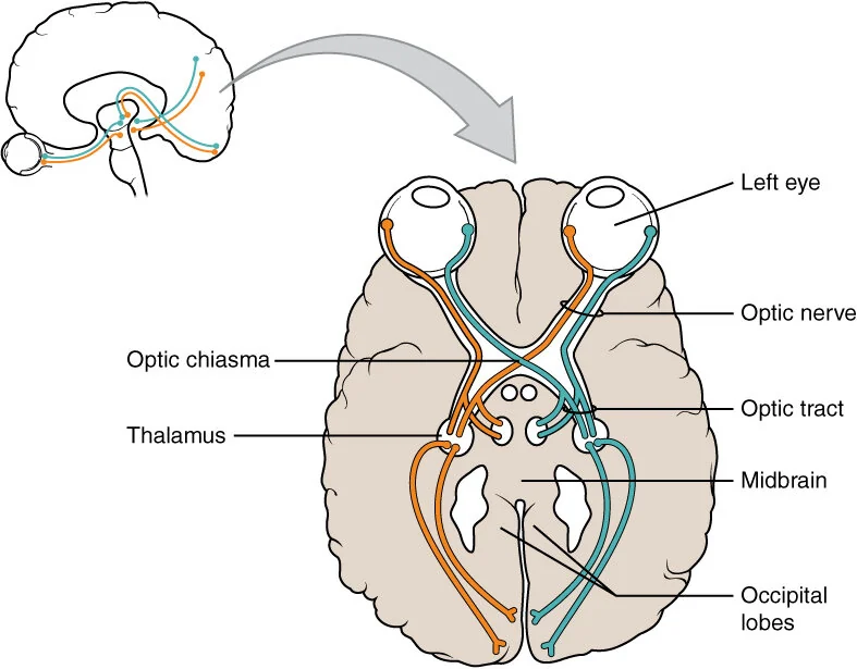
eye → optic nerve → optic chiasm → optic tract → thalamus (lateral geniculate nucleus) → occipital lobe, where area V1 sits
OpenStax · Anatomy and Physiology (2016) · CC BY 4.0
How far this holds
The structures and the numbers are well established, but they were measured outside an interface. The jump from here to the screen is mine.
The numbers for the visual pathway come from recordings in primate cortex and from functional imaging in fixation tasks: dots on a black screen, not product screens. And the focus isn’t strictly single. In tasks of tracking moving targets, attention splits into as many as four, with a separate limit for each half of the visual field, as AlvarezOne reference in this work:2005Independent resources for attentional tracking in the left and right visual hemifieldsSee in the bibliography → and Cavanagh measured in 2005. The cost of splitting shows up in precision, not in cancellation.
The bottleneck belongs to the system, not to the person, and it isn’t the same in everyone: someone with low vision, a reduced visual field or a screen reader doesn’t receive the whole scene and then filter it, they receive it already in series, one piece at a time. Much of what this chapter calls “visual attention” becomes, there, reading order and structure of the markup. The limit still holds, only it bites in the reading order and in the markup, and not in the position on the screen.
Hearing
Several sounds arrive at the same time; only one becomes listening.
Sound takes a similar path. It enters through the ears, goes down the cochlea and activates the hair cells; the impulse continues to the cochlear nucleus, in the brainstemThe oldest part of the brain, at the junction with the spinal cord. It handles what is non-negotiable — breathing, waking, reacting to a fright — and it is where almost all the signals from the senses pass through.See it in Senses and attention →See in the glossary →, then to the inferior superior colliculusA station in the brainstem that pulls the gaze towards what moves or contrasts, before the person decides to look. It is that reflex that every visual highlight hijacks.See in the glossary →, in the midbrain, to the medial thalamusThe brain’s distribution hub: almost everything that comes from the senses passes through it before reaching the cortex. Smell is the odd one out, with a direct route to the areas of emotion and memory, and that is where the feeling-laden memory a scent pulls up comes from.See it in Senses and attention →See in the glossary → body, in the thalamus, and finally to the A1The first area to receive sound, in the temporal lobe. It receives everything that comes in through the ears, but only one stream at a time gets priority and reaches consciousness.See it in Senses and attention →See in the glossary →, area A1, in the temporal lobe.
What happens when several sounds arrive at the same time was measured in 1953, in the experiment that became known as the cocktail party effect: Colin CherryRan the cocktail party experiment: how the ear picks one voice among several and what happens to the ones left out.One reference in this work:1953Some experiments on the recognition of speech, with one and with two earsSee on Wikipedia ↗See in the bibliography → showed that, among several voices, we choose only one to hear consciously. Magnetoencephalography later confirmed that attention already amplifies the chosen stream inside the auditory cortex itself, between 20 and 50 milliseconds after the sound, before any conscious decision, as WoldorffMeasured that auditory attention amplifies the signal as early as the first 20 to 50 milliseconds, before any conscious decision.One reference in this work:1993Modulation of early sensory processing in human auditory cortex during auditory selective…See in the bibliography → and colleagues recorded in 1993. It’s this resolution in milliseconds that magnetoencephalography and EEG deliver, and that no other method delivers. The range the ear hears is old too. From the fossilised ossicles of five individuals from Atapuerca, MartínezMeasured the inner ear of 350,000-year-old fossils and showed that the human hearing range was already ours.One reference in this work:2004Auditory capacities in Middle Pleistocene humans from the Sierra de Atapuerca in SpainSee in the bibliography → and colleagues reconstructed in 2004 the transmission of sound through the outer and middle ear, and it already followed the human pattern in the middle range of speech, 2–4 kHz, and not the chimpanzee’s. In the 3.5–5 kHz range the picture is different: there the ones from Atapuerca were not yet modern, and the ones who get there are the Neanderthals, as Conde-ValverdeOne reference in this work:2021Neanderthals and Homo sapiens had similar auditory and speech capacitiesSee in the bibliography → and colleagues showed in 2021. The fossils were dated again after publication and are at least 430,000 years old, older than our own species. However many audio channels an app offers, we remain limited to a single conscious channel.
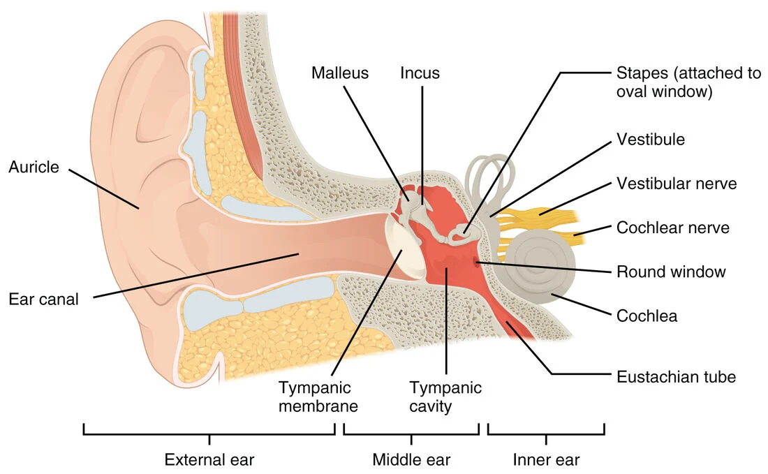
cochlea → vestibulocochlear nerve → cochlear nucleus → inferior colliculus → thalamus (medial geniculate nucleus) → primary auditory cortex (A1), in the temporal lobe
OpenStax · Anatomy and Physiology (2016) · CC BY 4.0
How far this holds
What this part claims was measured this way, and the studies are in the bibliography.
Colin CherryRan the cocktail party experiment: how the ear picks one voice among several and what happens to the ones left out.One reference in this work:1953Some experiments on the recognition of speech, with one and with two earsSee on Wikipedia ↗See in the bibliography → measured with headphones and two simultaneous voices, asking the person to repeat one of them out loud. The Atapuerca ossicles were reconstructed by tomography and acoustic modelling. Neither is an interface, but both measure what this part claims.
Touch and proprioception
The hand that operates your interface is, in the brain, bigger than the entire back.
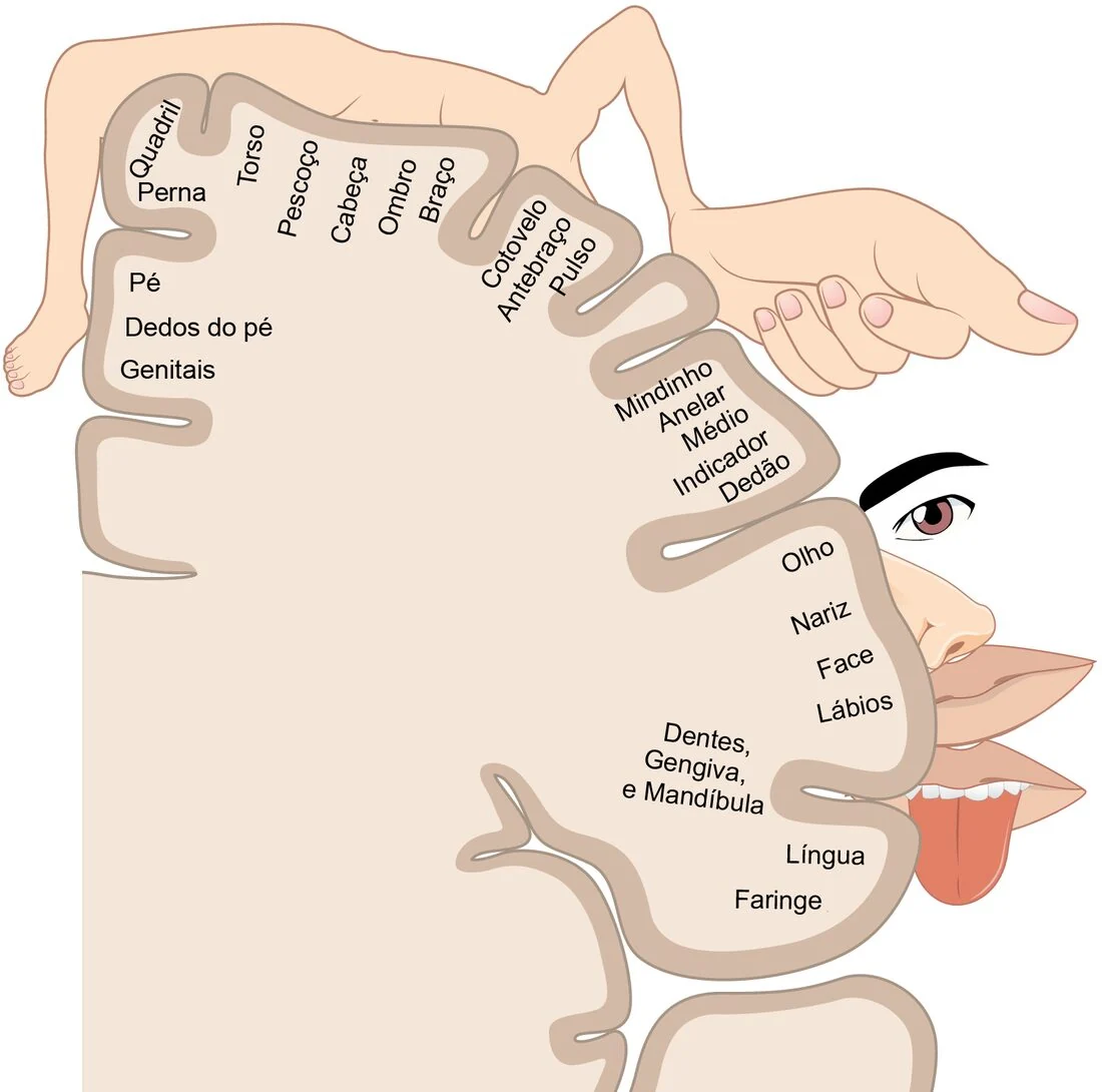
Touch enters through the skin and climbs. When we touch something, mechanical receptors generate impulses that reach the dorsal root ganglion and travel, through the spinal cord, to the thalamusThe brain’s distribution hub: almost everything that comes from the senses passes through it before reaching the cortex. Smell is the odd one out, with a direct route to the areas of emotion and memory, and that is where the feeling-laden memory a scent pulls up comes from.See it in Senses and attention →See in the glossary → and to the S1The map of the body drawn over the cortex. Fingers, lips and tongue take up enormous areas; back and legs, almost nothing: that is why the fingertip tells apart what the elbow cannot.See it in Senses and attention →See in the glossary →, S1, in the postcentral gyrus. There are four types of receptor, Merkel discs, Meissner corpuscles, Ruffini endings and Pacinian corpuscles, and the map they form in S1 is a mirror of the body’s surface, only a distorted mirror: hand and lips take up a disproportionate slice of the cortex, while the whole trunk fits into a small patch. That’s how Wilder PenfieldStimulated the cortex of awake patients during surgery and drew the homunculus, the body as the brain represents it.One reference in this work:1937Somatic motor and sensory representation in the cerebral cortex of manSee on Wikipedia ↗See in the bibliography → and Boldrey drew the homunculus, in 1937, by directly stimulating the cortex of awake patients.
The same disproportion shows up in the skin, when you count. The hairless hand, palm and fingers, has about 17,000 mechanoreceptive units, and they aren’t evenly distributed: at the fingertip the density reaches about 240 per square centimetre, against about 60 on the palm, in the count JohanssonCounted, with Vallbo, the mechanoreceptive units of the human hand one by one, and measured the density of each type in each region of the glabrous skin.One reference in this work:1979Tactile sensibility in the human hand: relative and absolute densities of four types of me…See in the bibliography → and Vallbo made in 1979 recording fibre by fibre. Pacinian corpuscles, the type that responds to vibration, have their peak sensitivity around 250 Hz, and that’s why phone haptic motors are built to resonate between 175 and 235 Hz. The range didn’t come from an engineering choice. It’s the frequency the body feels best.
OpenStax College; derivative work by Ederporto · Anatomy & Physiology (Portuguese edition) · CC BY 3.0
How far this holds
What this part claims was measured this way, and the studies are in the bibliography.
Wilder PenfieldStimulated the cortex of awake patients during surgery and drew the homunculus, the body as the brain represents it.One reference in this work:1937Somatic motor and sensory representation in the cerebral cortex of manSee on Wikipedia ↗See in the bibliography → stimulated the cortex of awake patients during surgery and noted down what each one reported feeling. JohanssonCounted, with Vallbo, the mechanoreceptive units of the human hand one by one, and measured the density of each type in each region of the glabrous skin.One reference in this work:1979Tactile sensibility in the human hand: relative and absolute densities of four types of me…See in the bibliography → and Vallbo counted the units by recording fibre by fibre in volunteers, with a microelectrode in the nerve of the arm.
Smell and taste
The only sense with a direct shortcut to emotion, and the one with the lowest bandwidth of all.
Smell and flavour enter through narrower doors. Smells are picked up by receptor cells in the olfactory mucosa and sent directly to the olfactory bulb, which projects to the piriform cortex and to limbic structures such as the amygdalaTwo small structures in the temporal lobe that decide, in milliseconds, whether something is a threat or an opportunity. They respond to your screen before any conscious judgement about it.See it in Emotion →See in the glossary → and the hippocampusThe structure that turns recent experience into lasting memory. Everything your interface asks the person to remember tomorrow passes through it today.See it in Memory →See in the glossary →. That’s why certain aromas immediately evoke an emotion or a memory: smell is the only sense that reaches the limbic systemAn inherited name for a group of structures — amygdala, hippocampus, neighbours — that the design literature still treats as a single block. Neuroscience does not treat it that way, because there is no anatomical boundary and no common function, and each of these structures does different things.See it in Emotion →See in the glossary → at the very first stop, without passing through the thalamusThe brain’s distribution hub: almost everything that comes from the senses passes through it before reaching the cortex. Smell is the odd one out, with a direct route to the areas of emotion and memory, and that is where the feeling-laden memory a scent pulls up comes from.See it in Senses and attention →See in the glossary → first. Even with about half of our olfactory receptor genes inactive, we keep something around 400 functional ones, in the count MalnicOne reference in this work:2004The human olfactory receptor gene familySee in the bibliography →, Godfrey and Buck published in 2004.
Taste depends on the taste buds, which transmit sweet, salty, sour, bitter and umami to the solitary tract, in the brainstemThe oldest part of the brain, at the junction with the spinal cord. It handles what is non-negotiable — breathing, waking, reacting to a fright — and it is where almost all the signals from the senses pass through.See it in Senses and attention →See in the glossary →, from where the signals rise to the thalamus and reach the gustatory cortex, in the insular lobe. The resolution of smells and flavours remains low, and not even the structure of a molecule predicts well what it will smell like, as KellerMeasured the perception of chemically diverse molecules and showed how much the human sense of smell discriminates, and how much it fails to describe.One reference in this work:2016Olfactory perception of chemically diverse moleculesSee in the bibliography → and Vosshall measured in 2016 with hundreds of compounds. Describing a smell in detail is nearly impossible, and what fails there is naming, not memory, as OlofssonExplained why it is so hard to name a smell: the sense of smell has a weak link to the language system.One reference in this work:2015The muted sense: neurocognitive limitations of olfactory languageSee in the bibliography → and Gottfried showed in 2015. Any attempt to digitise odours runs into a bandwidth far smaller than that of video or audio.
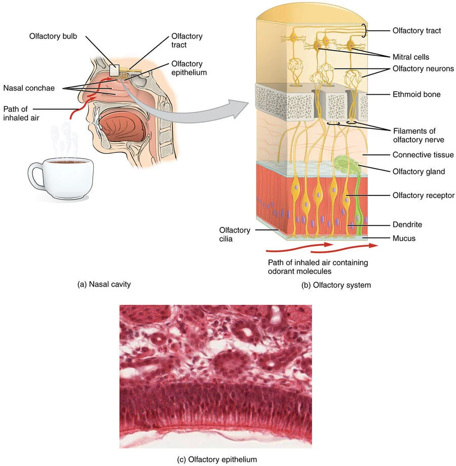
olfactory epithelium → olfactory bulb → piriform cortex → amygdala and hippocampus. It is the only sense that reaches the limbic system at the very first stop, without passing through the thalamus first
OpenStax · Anatomy and Physiology (2016) · CC BY 4.0
How far this holds
What this part claims was measured this way, and the studies are in the bibliography.
Counts of receptor genes by sequencing, and detection thresholds measured in a booth, with vials. The part about describing a smell comes from naming tests: the person sniffs and tries to say what it is.
This part is the shortest on the page, and on purpose: it’s the sense with the lowest bandwidth and it’s also the least studied of the five. The line that circulates — that smell has a “direct line to emotion” — is true in the anatomical wiring and tends to be stretched too far in the conclusion. Reaching the limbic systemAn inherited name for a group of structures — amygdala, hippocampus, neighbours — that the design literature still treats as a single block. Neuroscience does not treat it that way, because there is no anatomical boundary and no common function, and each of these structures does different things.See it in Emotion →See in the glossary → first doesn’t mean producing a stronger or more reliable emotion, and there is no interface application that depends on it.
Vestibular system
The sensor that makes you sick in the car is never switched off.
To keep our balance and perceive movement, we rely on the semicircular canalsThree rings of fluid in the inner ear, one for each axis of rotation. When the head turns, the fluid takes a moment to follow, and it is that delay the body reads as “I turned”.See it in Senses and attention →See in the glossary → and the otolithsTwo pouches with calcium crystals on a membrane, also in the inner ear. They register acceleration in a straight line and the direction of gravity, which way is down.See it in Senses and attention →See in the glossary → in the labyrinth of the inner ear. The first register rotation; the second, straight-line displacement and the direction of gravity. Both send signals to the vestibular nucleus in the brainstemThe oldest part of the brain, at the junction with the spinal cord. It handles what is non-negotiable — breathing, waking, reacting to a fright — and it is where almost all the signals from the senses pass through.See it in Senses and attention →See in the glossary → and, from there, to the cerebellumThe structure low at the back of the skull that automates movement and sequence. The more someone uses your product, the more the operation migrates here, and the costlier it gets to change what has already become automatic.See it in Memory →See in the glossary → and the vestibular cortex.
And there’s something here with no parallel in the other senses: they fire all the time. Even with the head still, a semicircular canal fibre keeps up about ninety impulses per second. There is no off position. Movement doesn’t make the signal appear. It makes that rate rise on one side of the head and fall on the other.
The brain is constantly comparing what the eye reports with what the inner ear reports, and it counts on the two saying the same thing. When they disagree, you get sick. And the two ways of making them disagree are symmetrical. In the car, looking at your phone, the ear says you’re moving and the eye sees a page standing still. In a virtual reality headset, sitting down, the eye sees you walking down a corridor and the ear says nobody has moved. It’s the same disagreement, with the roles swapped, and that’s why looking out of the side window helps in one case and closing your eyes helps in the other.
On a screen, what produces the disagreement is delay: you turn your head, the inner ear registers it instantly, and the image arrives a few moments later. Note that the delay isn’t between one sense and another. It’s between the movement and the drawing, and it’s the late arrival of the drawing that makes the two senses tell different stories.
It’s worth noticing where the numbers that get repeated about this come from, because they are a textbook case of a number changing owners along the way. Laviola Jr.Reviewed what is known about sickness in virtual environments, the mismatch between what the eye sees and what the inner ear feels, and what it costs whoever uses them.One reference in this work:2000A discussion of cybersickness in virtual environmentsSee in the bibliography → describes delay as a cause, in 2000, and gives no threshold at all: in the section on delay there isn’t a single millisecond, and the display number he does give is 30 Hz, about flicker, which is another problem. Thirty hertz is the common video rate, and flicker is that shimmer a fluorescent lamp makes in the corner of your eye.
The famous “under 20 milliseconds” is a design recommendation John Carmack made in 2013, and it became physiological fact along the way. For a ruler: at 60 frames per second, each frame lasts 16.7 ms. In other words, the recommendation is “delay shorter than one frame”, something anyone who has ever tuned an animation knows by the name of dropped frame.
What was actually measured is something else. People detect delay below 17 ms, and one participant detected 3.2 ms; the studies that induced sickness on purpose, meanwhile, worked with delays from a few tens of milliseconds up to 340 ms, in the review StauffertReviewed what was actually measured about latency and sickness in virtual reality, and separated what is a measured threshold from what is a design recommendation turned into fact.One reference in this work:2020Latency and cybersickness: impact, causes, and measuresSee in the bibliography → and colleagues published in 2020. That is, between two and twenty frames. Detecting isn’t getting sick, and the two numbers keep being swapped for one another, even though the two ranges touch at the bottom end.
This seemed to be the rare case where permanence can be measured in fossils, because the bony labyrinth is bone and bone survives. I went and checked, and the finding says otherwise: the labyrinth of the Atapuerca hominins has a derived pattern of semicircular canal proportions, characteristic of the Neanderthal lineage, and not the modern geometry, as QuamReconstructed the bony labyrinth of the Atapuerca hominins and found a derived pattern, of the Neanderthal lineage, not the modern human geometry.One reference in this work:2016The bony labyrinth of the middle Pleistocene Sima de los Huesos hominins (Sierra de Atapue…See in the bibliography → and colleagues described in 2016. So this sensor doesn’t serve as an example of “the same for hundreds of thousands of years”. The one that does is the ear, whose fossil ossicles already transmitted sound the way ours do.
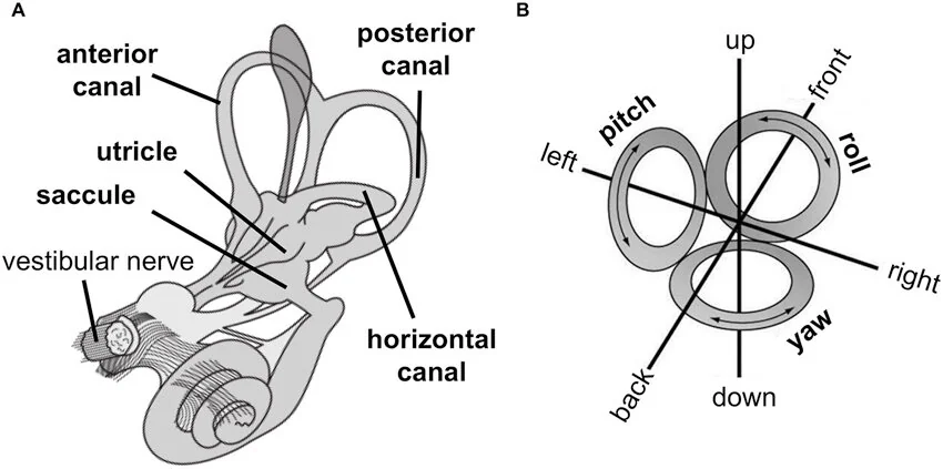
semicircular canals (rotation) and utricle and saccule (linear acceleration) → vestibular nerve → vestibular nuclei → cerebellum and vestibular cortex
Christian Pfeiffer, Andrea Serino and Olaf Blanke · Frontiers in Integrative Neuroscience (2014) · CC BY 3.0
How far this holds
The structures and the numbers are well established, but they were measured outside an interface. The jump from here to the screen is mine.
The firing rates come from recordings in the vestibular nerve of primates, with the head under controlled rotation in a chair. Virtual reality sickness was measured separately, in another literature and with another method.
The other parts of the engine